Tuesday, November 27, 2018

More random images

As the US-based team make their final preparations to depart Nepal, we leave you with yet another collection of random images taken during our time here. We again sincerely thank the Jirel community for their wonderful support for making this project a huge success. We also thank you for your interest. We'll be in touch soon with more blog updates. Until then...


Mascot: Team JES.


Issues with the slit lamp.

Months of the year, written and numbered.

A slit lamp being borrowed from the Jiri Community Eye Center (CEC).

View from the clinic between centrifuge spins.

Jiri school bus.

Lunch dining area.

Sarangi: a folk Nepalese bowed string instrument.


Sunset, from the clinic.

The front gates to the clinic.

Snow capped mountains, in the background.

More of the snow capped mountains, on the drive back to Kathmandu.

A full moon peaking over the Jiri landscape.

Time to get more of this...

Sunday, November 25, 2018

The Jiri Eye Study: By the numbers

While every field trip to collect data is a busy, intense period, this trip has been a little busier than usual. In addition to collecting data, the investigative team have also submitted a couple of grant applications to the NIH (National Institutes of Health), worked on a couple of abstracts that will soon be submitted for presentation at the 2019 ARVO (Association for Research in Vision and Ophthalmology) annual meeting, and worked on revising a manuscript (after reviewer comments) that reports the genetics of glycated serum protein, a potential biomarker for glycemia, in the Jirel pedigree. Therefore, we'll have time to report on some of the interesting eye pathology observed on this trip in the coming weeks.

As the sun here in Jiri is set to rise, the research team are starting to make their final preparations for the road trip back to Kathmandu. In the meantime, we thought we'd give you a breakdown of the data collection activities here in Jiri;

3: The number of slit lamps used.
4: The number of years taken to collect the data.
8: The number of field trips to Jiri.
12: The number of sampling days per field trip.
16: The number of weeks spent in Jiri.
21: The average number of Jirels seen per day, per field trip.
96: The total number of sampling days.
255: The average number of Jirels seen per field trip.
510: The average number of Jirels seen per year.
1,123: The approximate number of Jirel females (~55%) recruited into the study.
2,042: The total number of Jirels recruited into the Jiri Eye Study - that's 102.1% of our recruitment target!
4,000: The approximate number of transfer pipettes used.
44,924: The approximate number of pages of data scanned.
180,016: Approximate number of miles traveled by the US-based team.
290,944: Approximate number of kilometers traveled by the US-based team.


Friday, November 23, 2018

SAMPLE SIZE: n = 2,000!!


Today marked a major milestone for the Jiri Eye Study; we reached our recruitment goal of 2,000 members of the Jirel population! In fact, we finished the day at 2,003 individuals and we still have two more days of sampling to go.

This milestone is a major achievement, and is testament to the hard working and dedicated members of the field work team from the University of Texas Rio Grande Valley, Tilganga Institute of Ophthalmology, Miami University, and Wright State University.

On behalf of the research team, we sincerely thank the Jirel people for their cooperation with this important project – investigating the genetic epidemiology of ocular health and disease. Additionally, we thank the Jirel people for their continued support on all the research projects that have been conducted in the region over the past 30 years.

The Jiri Eye Study field work team and the 2,000th research participant and his wife, the 2,001st participant
The photo was taken with the permission of both participants.


Monday, November 12, 2018

Hello, Jiri, it's been a while...

In fact, it's been a while for all, actually. While the team has been busy with grant applications, we are now back in Jiri and looking forward to another successful field trip to collect more data and continue to improve the ocular health of the Jirel community. On this trip we expect to pass another great milestone so stay tuned for more in the coming days. In the meantime, here's a couple of pictures of home for the next couple of weeks...


Monday, March 26, 2018

Random images from Jiri, Nepal

As the US-based team bids farewell to our Nepali friends/colleagues from Tilganga and gets ready to depart Kathmandu for home, we leave you with yet another collection of random images taken throughout our time here, this time from Jiri. We sincerely thank the Jirel community and thank you for your interest. We'll be in touch very soon with more blog updates. Until then...


Sunday, March 25, 2018

A safe return to Kathmandu; n = 1,801!

The research team is now back safe and resting up in Kathmandu after yet another successful field trip. We again exceeded our goal of 250 individuals with 254 individuals recruited to the project. This now brings the total recruitment to 1,801 individuals who have completed all elements of the comprehensive eye exam and other aspects of data collection. We have a few more official duties at the Tilganga Institute of Ophthalmology today before we start to prepare for our journey home tomorrow. In the meantime, here's a brief report on our journey from Jiri to Kathmandu...

We left Jiri a bit before 08:00 h, one of our earliest and successful departures to date.

Sunrise, the morning of our departure from Jiri.

But it wasn't long before we made one of several stops along the way...

#1: Toilet break for Dr. Suman-ji's pet dog.


This first stop did allow us to capture a picture of Chhettrappa (right of picture), though, the village where most of the Jirels recruited on this trip reside.

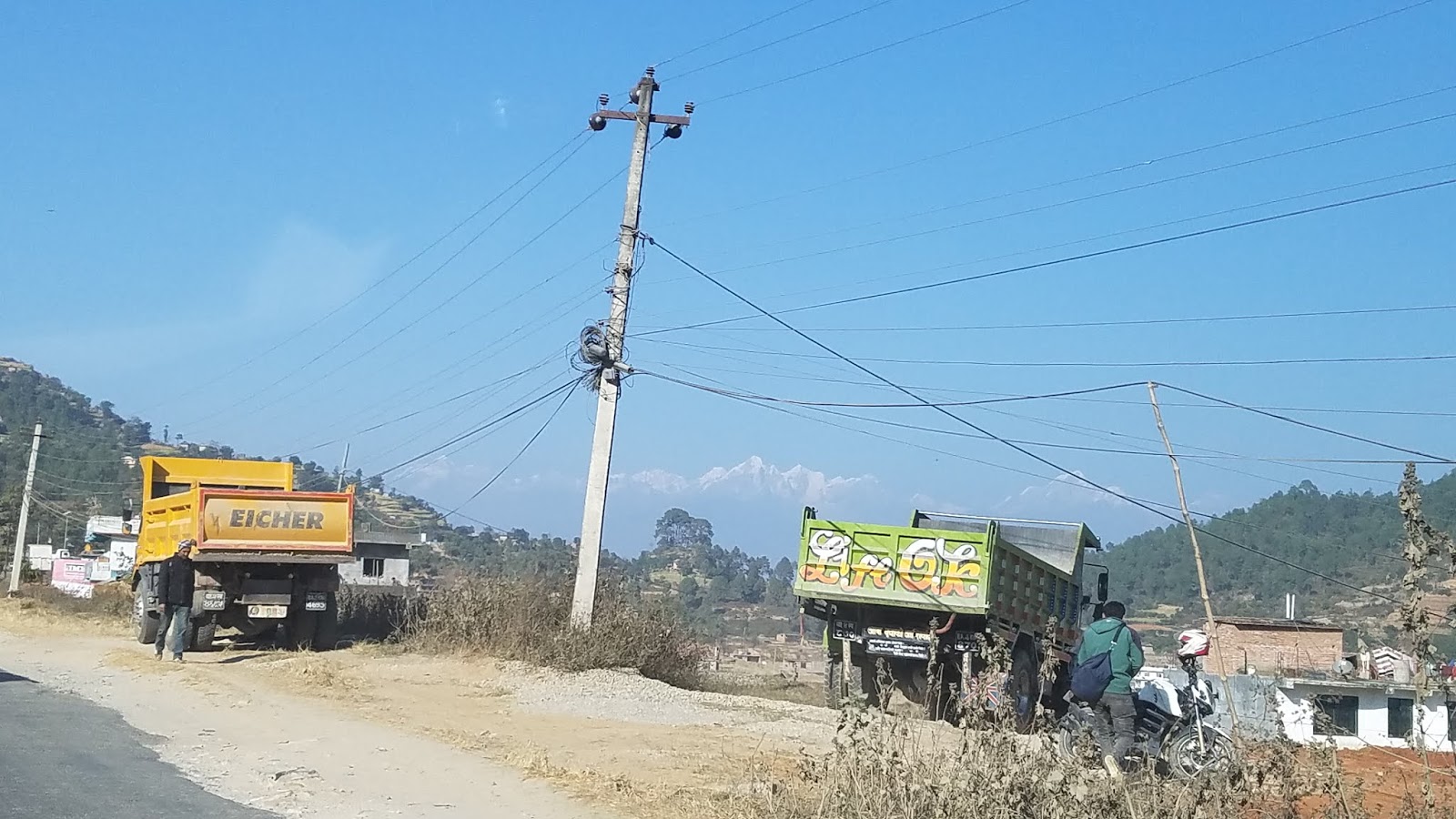
#2: We stop to find a truck, stopped along a temporary one lane section of the road, being loaded with rocks. Fortunately, we didn't have to wait too long as the truck was near fully loaded.


#3: Through Rammechap, the road was blocked in both directions as a backhoe loader was lifting what appeared to be a large, heavy printing press.


Not long after this third stop, as we negotiated the traffic backlog...


#4: ...we come to a fourth stop. Another truck was being loaded with dirt and this time it was the backhoe loader that was obstructing our path.


#5: Once we had some open road we had to slow down for things like...steamrollers and tractors pulling trailers!


#6: Until we came to another complete stop to allow police officers to direct the flow of traffic along a temporary one lane section.


And all of this was before lunch!! After which, we enjoyed a good run home.


Only to hit the customary traffic congestion in Kathmandu.


Wednesday, March 21, 2018

Macular hole

In what has been observed in a few of the JES research participants to date, is what is known as a macular hole. As part of the normal aging process, the vitreous humor (also termed, vitreous) will slowly contract and pull away from the surface of the retina. However, if the vitreous is firmly attached, or sticks to the retina when it pulls away, this can cause the macula to stretch and create a tear in the retina, thus creating a macular hole. As the hole forms, one's central vision will become blurred or distorted (remembering that the macula, the central part of the retina, is responsible for central, high-resolution vision). As the hole grows, a dark or blind spot will appear in the central vision. Peripheral vision is not affected by a macular hole.

In the fundus (red and red free) image below, a macular hole is observed. The macula (the dark 'spot' in the near center of the fundus image) can best be described by having a clear and defined boundary.


In comparison, in the fundus image (below) of a normal eye, the macula is best described by having a more diffuse (less defined) boundary.


With the aid of OCT technology, a macular hole is more clearly displayed. In the left image (below), the macular hole is clearly evident when compared to a normal, healthy retina (right image below). Both OCT images of the retina are captured traversing the superior-inferior quadrants of the retina.

Friday, March 16, 2018

Diabetes, hypertension and ocular health

Diabetes and hypertension (high blood pressure) are systemic diseases that inflict a significant impact on global health. One of the many complications arising from these systemic diseases is perturbed ocular health, for example, retinal hemorrhageHypertensive retinopathy and diabetic retinopathy are two such conditions where retinal hemorrhages can occur.

Here, we see Dr. Suman-ji (ophthalmologist) and Pradeep-ji (OCT specialist) review fundus photos at the conclusion of a day's sampling.


Following the review, one research participant had what appeared to be a retinal hemorrhage. In the participants fundus photograph (below), the hemorrhage (red 'spot') can be seen below the macula (dark 'spot' just left of center) and at about '8 o'clock' to the optic nerve head (bright 'spot' to the right of the image). While the participant did have high blood pressure (systolic and diastolic blood pressure > 140 and 90 mm Hg, respectively), we cannot definitely conclude that this high blood pressure is the cause of the hemorrhage.